Univ.-Prof. Dr. Lutz Nasdala
Stellv. Institutsvorstand
lutz.nasdala(at)univie.ac.at
Josef-Holaubek-Platz 2 (UZA II),
1090 Wien
Raumnummer: 2A251
T: +43-1-4277-53220

Research interests and current co-operation and activities
My main research interests lie in the general area of mineralogy and mineral physics. They are particularly related to the study of real structures of minerals (i.e., deviations from the ideal structural and chemical composition) and their use for the interpretation of mineral and rock formation and alteration processes. They include the following areas in which I plan to continue my research in the next years (selection, not complete):
(a) Radiation damage in U-Th-bearing minerals
Structural determination, and characterization of the variations in the chemical and physical behavior, such as changes in the diffusion of helium. The main research goal is to contribute to the understanding of effects on radiometric age determinations and potential immobilization and alteration processes in radioactive waste forms, and to use the degree of damage retention to conclude on the thermal history of radioactive accessories and their host rocks.
Co-operation with P.W. Reiners (Tucson, AZ,), A.K. Kennedy (Perth, WA), R. Wirth (Potsdam), A. Kronz (Göttingen), S. Kostrovitsky (Irkutsk), Z. Losos, K. Sobek, R. Škoda (Brno), D.A. Zamyatin (Ekaterinburg), G.W.A.R. Fernando (Nugegoda), K.A.G. Sameera (Sri Jayawardenepura Kotte and Peradeniya), and others.
Cross-polarised transmitted light (a) and BSE image (b) of a heterogeneous zircon from the Saranac Prospect, Ontario. Finger-like altered areas (which are not affected by notable U leaching!) at the ends of fractures are recognised from their birefringence and dark BSE.
Images modified from Nasdala et al. (2010).

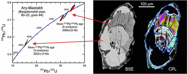
Back-scattered electrons (BSE) and cross-polarised light (CPL) images and Concordia plot of a zircon xenocryst from the Bargidamalah kimberlite pipe in Northern Yakutia. The main, ca. 2830 Ma old, U-rich interior region is metamict (see absence of any birefringence). The inherited, zoned core is lower in U and shows low to moderate interference colours, its U–Pb age is ca. 3570 Ma.
Images modified from Nasdala et al. (2014).
References:
Nasdala, L., Hanchar, J.M., Rhede, D., Kennedy, A.K., Váczi, T. (2010): Retention of uranium in complexly altered zircon: An example from Bancroft, Ontario. Chemical Geology, 269, 299-300.
Nasdala, L., Kostrovitsky, S., Kennedy, A.K., Zeug, M., Esenkulova, S.A. (2014): Retention of radiation damage in zircon xenocrysts from kimberlites, Northern Yakutia. Lithos, 206–207, 252–261.
Švecová, E., Copjaková, R., Losos, Z., Škoda, R., Nasdala, L. , Cícha, J. (2016): Multi-stage evolution of xenotime–(Y) from Písek pegmatites, Czech Republic: An electron probe micro-analysis and Raman spectroscopy study. Mineralogy & Petrology, 110, 747–765.
Zamyatin, D.A., Shchapova, Yu.V., Votyakov, S.L., Nasdala, L., Lenz, C. (2017): Alteration and chemical U-Th-total Pb dating of heterogeneous high-uranium zircon from a pegmatite from the Aduiskii Massif, Middle Urals, Russia. Mineralogy & Petrology, 111, 475–497.
Nasdala, L., Hauzenberger, C., Wildner, M., Chanmuang N., C. & Sameera, K.A.G. (2023): Metamict gadolinite–(Y) from Ratnapura, Sri Lanka. Journal of the Geological Society of Sri Lanka, 24(1), 23–30.
(b) Irradiation effects in minerals
Creation of structural changes in minerals and their synthetic analogues by light- and heavy-ion irradiation, in particular of FIB-prepared thin lamellae. Main goals include the understanding of the radiation-induced colouration (diamond, cordierite) and defect luminescence (zircon, monazite, quartz), and to contribute to a more quantitative estimation of structural damage caused by corpuscular irradiation.
Co-operation with S. Akhmadaliev (Dresden-Rossendorf), C. Chanmuang N., G. Habler (Wien), T. Váczi (Budapest), and others.
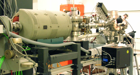
The nuclear reaction analysis beamline at the 5 MV tandem accelerator of the Forschungszentrum Dresden-Rossendorf, Germany, where ion-irradiation experiments were done.
Image courtesy T. Váczi.
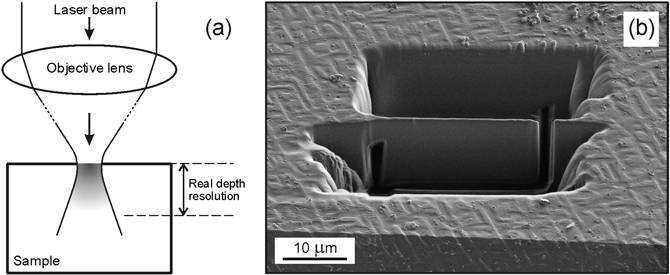
(a) Sketch visualising the depth resolution of a confocal spectrometer. (b) Preparation of thin samples in a focused ion beam (SE image). Conducting experiments with samples whose thicknesses are well below the spectrometer’s depth resolution performance helps a lot to exclude artefacts that may be caused by confocality limits.
Images modified from Nasdala et al. (2010).
Luminescence images of light-ion irradiated samples: (a) UV-excited PL image of a diamond slice irradiated along the direction of view. (b) OM-CL image of a synthetic zircon crystal (irradiation direction marked with arrow). In both cases, the ion irradiation has created defect-related emission centres.
Image (a) modified from Nasdala et al. (2013); image (b) modified from Nasdala et al. (2011).
References:
Nasdala, L., Grötzschel, R., Probst, S., Bleisteiner, B. (2010): Irradiation damage in monazite-(Ce): an example to establish the limits of Raman confocality and depth resolution. Canadian Mineralogist, 48, 351-359.
Nasdala, L., Grambole, D., Götze, J., Kempe, U., Váczi, T. (2011): Helium irradiation study on zircon. Contributions to Mineralogy and Petrology, 161, 777-789.
Nasdala, L., Grambole, D., Wildner, M., Gigler, A.M., Hainschwang, T., Zaitsev, A.M., Harris, J.W., Milledge, J., Schulze, D.J., Hofmeister, W., Balmer, W.A. (2013): Radio-colouration of diamond: a spectroscopic study. Contributions to Mineralogy and Petrology, 165, 843–861.
Schulze, D., Nasdala, L. (2016): Unusual paired pattern of radiohaloes on a diamond crystal from Guaniamo (Venezuela). Lithos, 265, 177–181.
Nasdala, L., Akhmadaliev, S., Burakov, B.E., Chanmuang N., C. & Škoda, R. (2020): The absence of metamictisation in natural monazite. Scientific Reports, 10, 14676.
(c) Experimental improvement and application of spectroscopic in situ-analyses
Identification of micrometre-sized phases and characterisation of their structural state, to contribute to the reconstruction of the samples’ formation and post-growth history. Study of absorption and emission centres in natural minerals with particular focus on REE centres in accessories.
Co-operation with J. Götze (Freiberg), M. Gaft ( Ariel), M. Wildner (Wien), V. Stähle, R. Altherr (Heidelberg), and others.
Study of suevite clasts contaning shock-induced veins, from the Ries impact crater. BSE images showing the occurrence of stishovite in sample ZLN124 (a) and jadeite in sample ZLN128 (b) along with Raman spectra and references (c).
Figures modified from Stähle et al. (2017).


Steady-state PL spectra of synthetic malayaite pigments (Ar+ 488 nm excitation), showing how effectively the presence of chromium quenches REE-related emissions.
Image modified from Nasdala et al. (2014).
References:
Pearson, D.G., Brenker, F.E., Nestola, F., McNeill, J., Nasdala, L., Hutchison, M.T., Matveev, S., Mather, K., Silversmit, G., Schmitz, S., Vekemans, B. Vincze, L. (2014): Hydrous mantle transition zone indicated by ringwoodite included within diamond. Nature, 507, 221–224.
Nasdala, L., Stoyanova Lyubenova, T., Gaft, M., Wildner, M., Diegor, W., Petautschnig, C., Talla, D. & Lenz, C. (2014): Photoluminescence of synthetic titanite-group pigments: A rare quenching effect. Chemie der Erde – Geochemistry, 74, 419–424.
Nasdala, L., Steger, S. , Reissner, C. (2016): Raman study of diamond-based abrasives, and possible artefacts in detecting UHP microdiamond. Lithos, 265, 317–327.
Stähle, V., Altherr, R., Nasdala, L., Trieloff, M., Varychev, A. (2017): Majoritic garnet grains within shock-induced melt veins in amphibolites from the Ries impact crater suggest ultrahigh crystallization pressures between 18 and 9 GPa. Contributions to Mineralogy and Petrology, 172, 86.
Nasdala, L. & Schmidt, C. (2020): Applications of Raman spectroscopy in mineralogy and geochemistry. Elements, 16(2), 99–104.
Zeug, M., Nasdala, L., Ende, M., Habler, G., Hauzenberger, C., Chanmuang N., C., Škoda, R., Topa, D., Wildner, M. & Wirth, R. (2021): The parisite–(Ce) enigma: challenges in the identification of fluorcarbonate minerals. Mineralogy and Petrology, 115, 1–19.
Hou, Z., Woś, D., Tschegg, C., Rogowitz, A., Rice, H.N., Nasdala, L., Fusseis, F., Szymczak, P. & Grasemann, B. (2023): 3D mineral dendrites reveal a non-classical crystallization pathway. Geology, 51, 626–630.
(d) Application of imaging and mapping techniques
Visualisation and characterisation of internal growth and alteration textures of minerals. Main goals include the physical understanding of the observed heterogeneity, investigation of artefacts as caused by the electron beam in the EPMA or the SEM, and the visualisation of patterns of internal stress / strain.
Co-operation with J. Götze (Freiberg), A. Kronz (Göttingen), T. Váczi (Budapest), R. Abart (Wien), and others.

In contrast to previous assumptions, the BSE intensity of natural zircon crystals was found not to be mainly controlled by the chemical composition: (a) Sketch of a heterogeneous zircon crystal that was cut in half, with one half being annealed to reconstitute the structure. (b) BSE image. (c) Colour-coded Raman map, visualising the degree of disorder. Note that the chemical zoning has rather mild effects on the observed BSE, whereas the BSE intensity correlates with the structural disorder (Raman FWHM broadening). In conclusion, the electron back-scatter intensity is strongly enhanced by structural damage.
Images modified from Nasdala et al. (2006).
Colour-coded EPMA element distribution images of an altered region adjacent to a large fracture in a fergusonite specimen from Madagascar. Uranium shows a rather chaotic distribution pattern, suggesting a non-uniform or even multi-step alteration progress. Yttrium is depleted in altered fergusonite areas compared to the unaltered bulk, but is a main constituent of the young fracture filling.
Images modified from Ruschel et al. (2010).
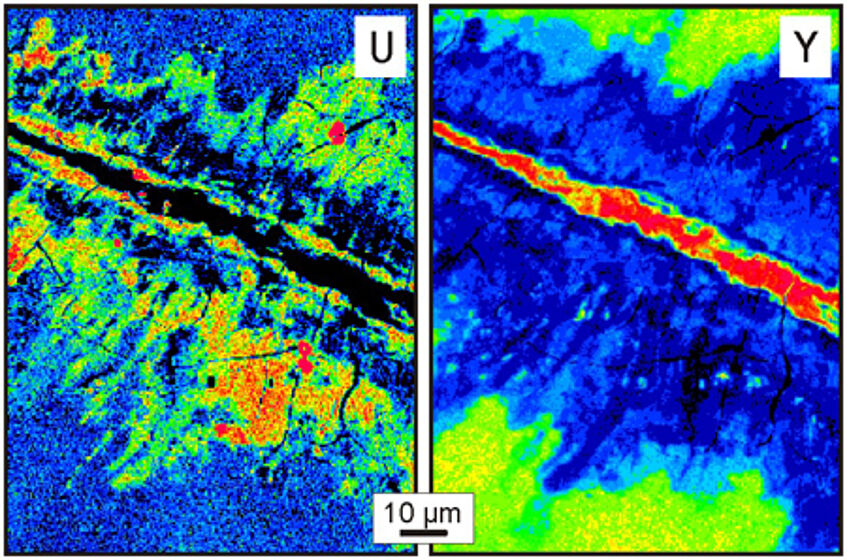
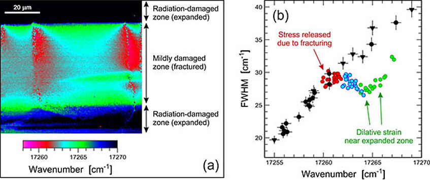
Photoluminescence (PL) study of a zoned zircon from Plešovice, Czech Republic. (a) Hyperspectral PL map, visualizing stress release near fractures (red). (b) Plot of FWHM against position of the Dy3+ emission line near 17,260 cm–1. Black, data for variably radiation-damaged, unstressed zircon (modified from Nasdala & Lenz, 2015).
References:
Nasdala, L., Kronz, A., Hanchar, J.M., Tichomirowa, M., Davis, D.D., Hofmeister, W. (2006): Effects of natural radiation damage on back-scattered electron images of single-crystals of minerals. American Mineralogist, 91, 1739-1746.
Ruschel, K., Nasdala, L., Rhede, D., Wirth, R., Lengauer, C.L., Libowitzky E. (2010): Chemical alteration patterns in metamict fergusonite. European Journal of Mineralogy, 22, 425-433.
Lenz, C., Nasdala, L. (2015): A photoluminescence study of REE3+ emissions in radiation-damaged zircon. American Mineralogist, 100, 1123–1133.
Nasdala., L. , Lenz, C. (2015): Photoluminescence spectroscopy: A tool for revealing stress caused by self-irradiation damage. Periodico di Mineralogia, 84, 127–128.
(e) Characterisation of gemstones
Study of gem materials (in particular with spectroscopic techniques) with regard to identification, formation and post-growth history (including treatment).
Co-operation with G. Giester, M. Wildner, C. Chanmuang N., C. Hauzenberger (Graz), R. Škoda (Brno), M.R. Scicchitano (Potsdam), B. Wanthanachaisaeng (Bangkok), E. Gamini Zoysa (Colombo), and others.
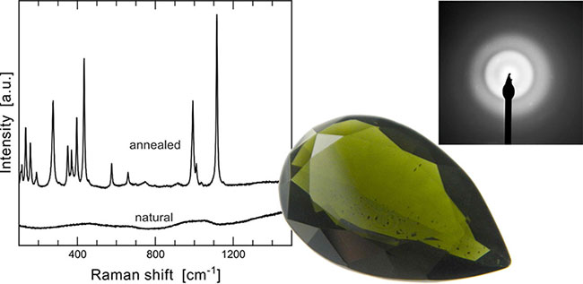
Facetted 7.05 ct "ekanite" from Eheliyagoda, Ratnapura, Sri Lanka. Left, Raman spectra before (amorphous) and after dry annealing (recrystallised). Right, electron diffraction pattern as obtained in the TEM, only showing "amorphous rings".
Images modified from Nasdala et al. (2017).
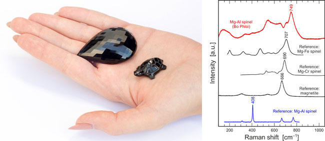
Droplet-shaped cut stone (167.4 ct) and carved turtle made of black spinel from Bo Phloi, Thailand. The Raman spectrum of the material differs from that of "normal" spinel, which is explained by the degree of inverse cation occupation.
Image courtesy M. Wildner.
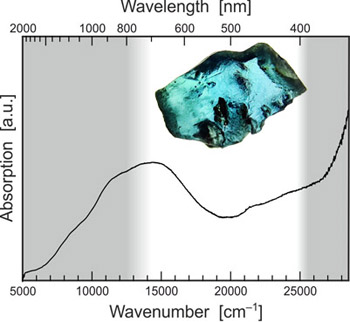
The blue colour of this rough serendibite (ca. 4.7 mm and 0.052 g) from Kolonna, Sri Lanka, is caused by an absorption band near 700 nm, assigned to Fe2+-Fe3+ intervalence charge transfer.
Image courtesy C. Chanmuang N.
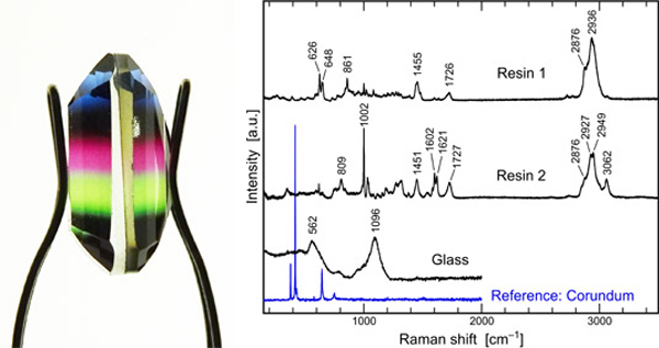
Glass doublet (6.7 ct). Shortly after the appearance of multicoloured synthetic corundum in the gem market (ca. 2019), the production and sale of cheap fakes increased. They consist of a coloured resin membrane sandwiched by pavilion and crown consisting of soda-lime glass.
References:
Zeug, M., Rodriguez Vargas, A.I. & Nasdala, L. (2017): Spectroscopic study of inclusions in gem corundum from Mercaderes, Cauca, Colombia. Physics and Chemistry of Minerals, 44, 221–233.
Nasdala, L., Corfu, F., Blaimauer, D., Chanmuang, C., Ruschel, K., Škoda, R., Wildner, M., Wirth, R., Zeug, M. & Zoysa, E.G. (2017): Neoproterozoic amorphous “ekanite” (Ca2Th0.9U0.1Si8O20) from Okkampitiya, Sri Lanka: A metamict gemstone with excellent lead-retention performance. Geology, 45, 919–922.
Zeug, M., Nasdala, L., Wanthanachaisaeng, B., Balmer, W.A., Corfu, F. & Wildner, M. (2018): Blue zircon from Ratanakiri, Cambodia. Journal of Gemmology, 36(2), 112–132.
Nasdala, L., Chanmuang N., C., Giester, G. & Wanthanachaisaeng, B. (2020): Multi-coloured synthetic corundum and multicoloured glass doublets in the Thai gem market. Journal of Gemmology, 37(1), 18–20.
Kruzslicz, Á.B., Nasdala, L., Wildner, M., Škoda, R., Redhammer, G.J., Hauzenberger, C. & Wanthanachaisaeng, B. (2020): Black spinel – a gem material from Bo Phloi, Thailand. Journal of Gemmology, 37(1), 66–79.
Chanmuang N., C., Nasdala, L., Wildner, M., Škoda, R. & Zoysa, E.G. (2021): Spectroscopic study of serendibite from Sri Lanka. Journal of Gemmology, 37(5), 451–454.
Wanthanachaisaeng, B., Nasdala, L., Chanmuang N., C. & Wildner, M. (2022): Quench-fractured bicoloured synthetic quartz: A doubly treated gem material. Journal of Gemmology, 38(2), 134–136.
Zeug, M., Nasdala, L., Chanmuang N., C. & Hauzenberger, C. (2022): Gem topaz from the Schneckenstein crag, Saxony, Germany: Mineralogical characterization and luminescence. Gems & Gemology, 58(1), 2-17.
Nasdala, L., Lamers, T., Gilg, H.A., Chanmuang N, C., Griesser, M., Kirchweger, F., Erlacher, A., Böhmler, M. & Giester, G. (2023): The Imperial Crown of the Holy Roman Empire, part I: Photoluminescence and Raman spectroscopic study of the gemstones. Journal of Gemmology, 38(5), 448–473.
(f) Proposing well-characterised minerals as analytical references
Retrieval and characterisation of highest-quality gem materials (especially zircon, titanite) in view of their possible use as international reference material for SIMS U–Pb geochronology (i.e. age determination using large-geometry ion microprobe systems) and stable-isotope analysis.
Co-operation with A.K. Kennedy (Perth, WA), P.W. Reiners (Tucson, AZ), F.-Y. Wu and Q.-L. Li (Beijing), J.W. Valley (Madison, WI), B. Wanthanachaisaeng (Bangkok), E.G. Zoysa (Mt. Lavinia, Sri Lanka), D. Szymanowski (Zürich), and others.
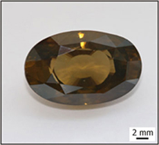
Zircon M257, a moderately radiation damaged but nevertheless extremely homogeneous, gem-quality specimen from Sri Lanka. Its chemical, isotopic, and structural composition has been characterised in detail. The 561.3 Ma old zircon is used as U–Pb reference material in the SIMS labs in Perth, Beijing, St. Petersburg, and Stockholm.
Image courtesy T. Häger.
Concordia plot of U-Pb data for the Sri Lankan reference zircon M127, obtained at the University of California at Santa Barbara (CA-TIMS; sixteen plateau steps) and at the University of Oslo (ID-TIMS, four individual analyses). All ellipses represent 2s uncertainties. The uncertainty of the mean age quoted includes uncertainties of decay constants.
Image from Nasdala et al. (2016).

References:
Nasdala, L., Hofmeister, W., Norberg, N., Mattinson, J. M., Corfu, F., Dörr, W., Kamo, S. L., Kennedy, A. K., Kronz, A., Reiners, P. W., Frei, D., Kosler, J., Wan, Y., Götze, J., Häger T., Kröner, A., Valley, J.W. (2008): Zircon M257 – a homogeneous natural reference material for the ion microprobe U-Pb analysis of zircon. Geostandards and Geoanalytical Research, 32, 247–265.
Kennedy, A.K., Kamo, S.L., Nasdala, L., Timms, N.E. (2010): Greenville skarn titanite: Potential reference material for SIMS U–Th–Pb analysis. Canadian Mineralogist, 48, 1423–1443.
Nasdala, L., Corfu, F., Valley, J.W., Spicuzza, M.J., Wu, F.-Y., Li, Q.-L., Yang, Y.-H., Fisher, C., Münker, C., Kennedy, A.K., Reiners, P.W., Kronz, A., Wiedenbeck, M., Wirth, R., Chanmuang, C., Zeug, M., Váczi, T., Norberg, N., Häger, T., Kröner, A. & Hofmeister, W. (2016): Zircon M127 – a reference material for U–Pb combined with Hf-, O-, and potentially Li-isotope analysis. Geostandards and Geoanalytical Research, 40, 457–475.
Szymanowski, D., Fehr, M.A., Guillong, M., Coble, M.A., Wotzlaw, J.-F., Nasdala, L., Ellis, B.S., Bachmann, O., Schönbichler, M. (2018): Isotope dilution anchoring of zircon reference materials for accurate Ti-in-zircon thermometry. Chemical Geology, 481, 146–154.
Nasdala, L., Corfu, F., Schoene, B., Tapster, S.R., Wall, J.W., Schmitz, M.D., Ovtcharova, M., Schaltegger, U., Kennedy, A.K., Kronz, A., Reiners, P.W., Yang., Y.-H., Wu, F.-Y., Gain, S.E.M., Griffin, W.L., Szymanowski, D., Chanmuang N., C., Ende, M., Valley, J.W., Spicuzza, M.J., Wanthanachaisaeng, B. & Giester, G . (2018): GZ7 and GZ8 – two zircon references for SIMS U–Pb geochronology. Geostandards and Geoanalytical Research 40, 457–475.